Ankle arthrodesis, more commonly known as ankle fusion, has remained a dependable surgical treatment for patients suffering from advanced arthritis, chronic instability, or severe post-traumatic deformity. While fusing the ankle means sacrificing joint motion, it offers patients the priceless relief of walking without pain. Among the available fixation methods, the Ankle nail system has gained significant preference in recent years. Its design allows solid fixation with minimal disruption to the surrounding soft tissues. Let’s look at how this procedure is carried out in a step-by-step fashion.
Surgical Steps for Ankle Arthrodesis
Planning Before Surgery
As with most orthopedic procedures, the real work begins well before the first incision. The surgeon thoroughly reviews the patient’s medical history and examines the overall alignment of the leg and foot. Standard X-rays are taken, and in complicated cases, CT or MRI scans may be necessary. A discussion with the patient is equally important at this stage—making sure they are aware of the expected outcome, which involves living without ankle motion but gaining a stable, pain-free gait.
Another vital part of planning is selecting the right nail. Length, diameter, and locking options must be matched to the patient’s anatomy. The surgical team also prepares bone graft material if gaps are likely to be encountered during the fusion.
Patient Setup and Anesthesia
Once inside the operating room, the patient is generally placed in a supine position. A radiolucent table is often used because it allows clear fluoroscopic imaging during surgery. Small adjustments such as a hip bump can help in bringing the foot into alignment. Depending on the case, regional anesthesia may be preferred for its postoperative pain benefits, though general anesthesia is also commonly used.
Accessing the Ankle
The approach does not require a large incision. A small cut is made, and careful dissection is carried out to reach the joint. Surgeons take great care at this point to avoid unnecessary damage to soft tissues, since preserving blood supply helps in healing.
Preparing the Joint
The key to a successful fusion is good preparation of the joint surfaces. The cartilage lining the tibia and talus is meticulously removed to expose raw bone. This ensures that once the nail is in place, the bones have a good chance to unite. The surgeon also focuses on alignment—keeping the ankle neutral in dorsiflexion with a slight degree of valgus, which positions the foot comfortably for walking. Bone grafts may be added if there are gaps or poor-quality bone.
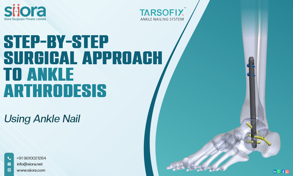
Placing the Nail
With the joint prepared, attention turns to nail insertion. A guidewire is first introduced through either the tibia or heel bone, depending on the chosen entry site. Correct placement is confirmed with live X-ray images. The canal is reamed to the required size, creating a pathway for the Ankle Nail.
The nail is then advanced slowly across the ankle joint, bridging the tibia, talus, and sometimes the calcaneus. Keeping the foot’s alignment correct during this step is critical—the nail essentially “locks in” the position chosen by the surgeon.
Locking the Nail
To complete fixation, locking screws are inserted proximally and distally using a targeting jig. This provides rotational stability and prevents any backing out of the nail. Some nail systems include compression options that apply pressure across the joint line, which significantly boosts the chance of successful fusion.
Closing the Wound and Recovery
After verifying the final alignment and nail position with fluoroscopy, the wound is thoroughly cleaned and closed in layers. A sterile dressing and cast or splint are applied.
For the first few weeks, patients are instructed to avoid weight-bearing to allow initial union. After around six to eight weeks, depending on healing progress seen in X-rays, gradual loading is introduced. Full fusion generally occurs after three to six months.
Why Surgeons Prefer the Ankle Nail?
- Smaller incisions and reduced soft-tissue damage compared to plate fixation
- Reliable stability thanks to its intramedullary, load-sharing design
- Suitable for patients with osteoporosis or deformity
- Faster procedures with predictable outcomes
Final Thoughts
Ankle arthrodesis using the Ankle Nail blends modern implant design with time-honored surgical principles. While the loss of ankle motion is inevitable, the procedure reliably delivers the stability and pain relief that patients with end-stage arthritis seek. Experienced surgical technique, thoughtful preoperative planning, and disciplined rehabilitation together ensure this method continues to stand as one of the most effective solutions in complex ankle pathology.
Learn more about the advancements in the healthcare industry, including the orthopedic sector at WHX Dubai 2026.
